daily health tips, health info, stay healthy every day, simple tips to improve your health, health and nutrition, health and wellness, healthy living, healthy diet, weight loss, beauty, simple health tips
Almost nobody knew her 20 years ago, and today more and more children suffer from chalk teeth - These are porous teeth where the enamel has not formed properly. The German Society for Dental, Oral and Maxillofacial Surgery warns of a new widespread disease.
When teeth just crumble away The molar-incisor-hypomineralisation - short MIH - affects according to recent studies about 10 to 15 percent of all children in Germany. Among the twelve-year-olds, more than 30 percent are affected by this disorder in the enamel. According to the German Oral Health Study, MIH is now more of a problem than dental caries. Cause still unknown The exact cause is still scientifically unclear, it is believed that it is probably an interaction of multiple triggers . Recent research indicates that the plasticizer bisphenol A might play a role in the formation of plastics. Other causes could include infectious diseases, antibiotics, chickenpox, environmental toxins such as dioxin, problems with pregnancy or upper respiratory tract disorders. The first year of life is crucial With MIH, incisors or milk teeth are less frequently affected. Most commonly, it shows on the molars, whose development occurs between the eighth month of pregnancy and the fourth year of life. This calcium and phosphate are stored, which harden the enamel. In the case of chalk teeth, the process is disrupted, leaving the enamel soft . In the worst case, the teeth are so porous that a part breaks off when penetrating the jaw. The first year of life seems to be decisive for the malformation. Subsequently, several factors seem to have come together for MIH to emerge. Fluorine can relieve symptoms Effective prevention of chalk teeth is currently not possible due to the lack of cause. According to experts, prophylaxis is all the more important because MIH teeth are particularly susceptible to caries. They therefore recommend fluoride in particular - in the form of toothpaste, fortified table salt, special varnish or mouth rinses. But above all for the thorough cleaning of the teeth to delay the almost inevitable caries as long as possible. If you notice any symptoms in you or your children, consult your dentist.
Risk factors - that contribute to the development of cardiovascular or congenital heart diseases are above all: unfavorable blood fat levels, high blood pressure, nicotine consumption, diabetes mellitus, overweight, lack of exercise and stress.
In addition to the causes that can be influenced by every human being, there are factors that are hardly or hardly influenceable, such as: familial predisposition age congenital heart defect. All of these risk factors may be involved in the development of cardiovascular disease or heart failure. 1. Unfavorable blood lipid levels These can be caused, among other things, by an unhealthy, one-sided diet with a high proportion of animal fats. Doctors also speak of LDL (Low Density Lipoprotein) 2. Hypertension When do you talk about hypertension? Blood pressure values below 120 mmHg systolic and 80 mmHg diastolic in an adult are considered optimal. Physicians only speak of high blood pressure (hypertension) when the blood pressure exceeds values of 140 mmHg systolic and 90 mmHg diastolic over a longer period of time. A classification of different degrees of severity of blood pressure can be found in the following table. 3. Nicotine consumption Smoking cigarettes is not only harmful to the lungs but also poison for the heart and blood vessels. Nicotine stimulates the heart to beat faster, increases the oxygen demand of the heart and has a narrowing effect on the vessels. The heart must therefore pump against increased resistance and thus provide increased performance to provide the body with sufficient oxygen and nutrients. In addition, smoking facilitates the formation of blood clots and thus increases the risk of thrombosis. Statistics show that smoking is a dangerous risk factor for heart attack, arteriosclerosis and stroke. For example, smokers suffer more heart attacks than non-smokers and often recover worse. Also, coronary heart disease occurs much more often among smokers. 4. Diabetes mellitus Diabetes mellitus (Greek for "honey-sweet flow" and popularly called "diabetes") is a chronic metabolic disorder. Glucose (simple sugar = glucose) is one of the carbohydrates and is the most important source of energy for the organism. Glucose is an important component of food and is obtained through the digestion and breakdown of carbohydrates eg starch (in cereals, potatoes, rice, fruit and fruit juices). Other important carbohydrate suppliers are cane sugar, lactose and the glycogen contained in the meat. Glucose enters the bloodstream via the intestinal wall. The blood supplies all cells with the energy donor glucose. The glucose concentration in the blood is normally 60-110 mg / dl. With increased energy requirements (e.g. sports, physical work), a body-own control system ensures that this level does not sink too much. With a higher carbohydrate intake, the glucose level rises temporarily. The lowering of the blood sugar level is mainly caused by insulin. Insulin is an endogenous hormone that is produced in the pancreas. In a healthy person, this hormone is released after eating carbohydrate-containing foods. It causes the cells to open their locks for sugar. There are different types of diabetes: Type 1 diabetes About half a million people have this type of diabetes. Cause of this disorder may be a lack of insulin secretion from the pancreas or a congenital or acquired insensitivity of the body cells against insulin (insulin resistance). Both result in insulin not being able to properly handle its transport pickup. symptoms:
extreme thirst
frequent urination
loss in weight
Type 2 diabetes This is a chronic disorder of carbohydrate metabolism . Due to a disorder of insulin secretion from the pancreas, insulin is missing after the meal. At the same time, insulin can not perform its transport task properly. This disorder is also due to a congenital or acquired insensitivity of the body cells to insulin (insulin resistance). If there is predominantly insulin deficiency in rather lean patients, this form is called type 2a diabetes. Here, the malfunction of the pancreas prevails in insulin production. Much more common, however, in about 6 million people in Germany, type 2b diabetes occurs. Most of these are affected by overweight people, where the insulin can not work properly - the glucose transport does not work. The glucose remains as too high blood sugar in the blood. In the long term, glucose is excreted by the kidneys, which is noticeable in the urine as uric acid. symptoms :
strong thirst
increased urination
blurred vision
cravings
susceptibility to infection
itching
badly healing wounds on the feet
5. Overweight Approximately 40% of Germans are too fat. If the person consumes more energy in the form of food than he actually consumes, he becomes overweight over time. Who wants to lose weight, must either eat less than he consumes or his energy consumption, eg. B. by sport or physical work significantly. Of obesity is when the weight is well above the normal weight. Reference value for the normal weight is, inter alia, the body mass index , which represents the body weight in relation to the body length. Consequences of overweight: metabolic disorders (diabetes, gout, high cholesterol). The risk of hypertension, heart attack, stroke, arteriosclerosis, fatty liver, thrombosis, joint disease, gallstones, increases significantly. What you can do: You should always take your time for the food. Chew well and do no other activities on the side. It is best to take five small meals a day. Always drink a large glass of liquid before eating, which reduces your appetite. With every diet you should drink a lot. Each diet should be accompanied by regular physical activity. The only sensible diet consists of a reduced-calorie mixed diet of a balanced diet with many fresh products. Unilateral diets are not recommended as they usually cause a lack of certain nutrients, vitamins or minerals and do not lead to a permanent change in dietary habits. Laxatives (e.g. with senna leaves), dehydrating agents (e.g. algae extract, birch leaves, pineapple enzyme) or detoxification stones (e.g. senna leaves and birch leaves) only lead to short-term, increased water excretion. In the long term important minerals can be lost. In addition, the regular intake of laxatives leads to constipation. A real weight reduction is not the case here. Important is a permanent change in diet , to avoid eating errors in the future and to keep the lower weight. 6. Lack of exercise An inactive lifestyle is a major factor in causing health problems. In particular, diabetes, back problems, overweight and cardiovascular diseases such as hypertension are consequences of lack of exercise. When physical activity is targeted, people of all ages can make a major contribution to preventing the development of disease and discomfort. For example, regular exercise reduces the risk of cardiovascular disease and reduces other risk factors of arteriosclerosis. Because sporting activities reduce blood pressure, weight and cholesterol levels. Sports such as Nordic walking, cycling, swimming, which are operated with moderation, ideally train the cardiovascular system. 7. Stress Under stress, stimuli are summarized that go beyond the individual perceived as normal level of physical and psychological stress. Detrimental stress (stress) is negatively experienced because the demands on oneself or those of the environment exceed their own abilities and possibilities. Stress factors (stressors) are, for example, pressure to perform, lack of time, lack of sleep, noise or changes in life circumstances such as puberty, divorce, unemployment or relocation. Excessive demands can lead to physical and mental illness symptoms. Because in stressful situations, the body releases more hormones that stimulate physical reactions. Among other things, they increase heart rate and blood pressure and dilate pupils and bronchi. If the stressors last longer, the body reaches a state of fatigue that can have different effects. Weight loss, sleep disorders, lack of concentration and depression are just a few of the consequences that can spread to high blood pressure, coronary heart disease, heart attack or asthma in the long term. Depending on the setting, stress is differentiated and perceived as follows: Positive stress A difficult situation is seen as a positive challenge that needs to be overcome. He expresses himself in high concentration and motivation and is the driving force for success. Negative stress A difficult situation is perceived as excessive demands. We feel at the mercy of the situation. There are no possibilities for action. In the long term, this negative stress makes you sick. Common causes Noise, sensory overload, mental stress, anger, disputes, illnesses, physical overexertion, performance, competition and time pressure, sleep deficit, events, e.g. marriage, birth, pregnancy, vacation It comes to the following body reactions: increased perception of the sensory organs, increased release of stress hormones such as adrenaline and norepinephrine, pulse rate, blood pressure and respiratory rate rise, sugar as an energy source is released, increased blood coagulation ability, lower blood flow to the digestive organs muscle tension. As a reaction to the stress hormones and the blood pressure change, the following symptoms may occur:
Congenital heart disease - is one of the most common anomalies of development and occupies third place after anomalies of the central nervous system and musculoskeletal system. The birth rate of children with congenital heart disease in all countries of the world ranges from 2.4 to 14.2 per 1,000 newborns. The incidence of congenital heart defects in live births is 0.7-1.2 per 1000 newborns. Errors with the same frequency of occurrence are often presented differently in the nosological structure in patients entering the cardiac departments (for example, a small defect of the interatrial septum and the tetralogy of Fallot). This is due to the varying degrees of threat to the health or life of the child.
The problems of diagnosis and treatment of congenital heart disease are extremely important in pediatric cardiology. As a rule, therapists and cardiologists are not sufficiently familiar with this pathology, as the vast majority of children are treated surgically or die without being adequately cared for at maturity. The causes of congenital heart disease are unclear. The most vulnerable period is 3-7 weeks gestation, that is, the time in which the heart structures are laid and formed. Of great importance are teratogenic factors of the environment, diseases of the mother and the father, infections, especially viral, as well as alcoholism of the parents, drug use, smoking of the mother. Many chromosomal diseases are associated with congenital heart defects. ICD code 10 Q20. Congenital anomalies (malformations) of the heart chambers and joints. Factors of survival in congenital heart disease Anatomo-morphological severity, i.e. type of pathology. The following prognostic groups are distinguished:
congenital heart defects with relatively favorable outcome: open arterial vein, interventricular septal defect (IVD), septal defect (SD), pulmonary artery stenosis; With these shortcomings, the natural mortality rate in the first year of life is 8-11%;
Fallot tetralogy, natural mortality in the first year of life - 24-36%;
complex congenital heart defects: left ventricular hypoplasia, pulmonary atresia, common arterial trunk; natural mortality in the first year of life - from 36-52% to 73-97%.
1.Age of the patient at the time of manifestation of the defect (the onset of clinical signs of hemodynamic disorders). 2.The presence of other (extracardiac) malformations increases mortality in one third of children with CHD to 90%. 3.Body weight at birth and premature birth. 4.Age of the child at the time of correction of the defect. 5.Severity and degree of hemodynamic changes, in particular - the degree of pulmonary hypertension. 6.Type and variant of cardiac surgery intervention.
The natural history of congenital heart disease Without surgical treatment, congenital heart defects occur in different ways. For example, in children 2-3 weeks hypoplastic left heart syndrome, atresia or pulmonary artery (with intact atrial septum) are rare in age, due to the high early-onset mortality in this vice. Overall mortality from congenital heart disease is high. At the end of the first week, 29% of newborns die in the first month - 42%, in the first year - 87% of children. Taking into account the modern possibilities of cardiosurgical care, it is possible to carry out the operation on a newborn in almost all congenital heart defects. However, not all children with congenital heart disease need surgery immediately after the pathology is revealed. Taking into account the tactics of treatment, patients with congenital heart disease are divided into three groups: 1.Patients in whom surgery for congenital heart disease is necessary and possible (about 52%); 2.Patients with no surgery due to minor hemodynamic disorders (approximately 31%); 3.Patients in whom correction of congenital heart disease is impossible and inoperable in the somatic state (about 17%). Before the doctor who conjectures congenital heart defect, there are the following tasks: Identification of symptoms ; ⦁Conducting a differential diagnosis with other diseases having similar clinical manifestations; ⦁the decision of a question on need of urgent advice of the expert (the cardiologist, the cardiologist); ⦁pathogenetic therapy. There are more than 90 variants of congenital heart disease and many of their combinations. Symptoms of a congenital heart disease When parents are interviewed, it is necessary to clarify the timing of the child's static functions: when he started to sit in the crib himself, he left. It is necessary to find out how the child gains weight in the first year of life, such as congestive heart failure and hypoxia, concomitant heart disease, accompanied by fatigue, "lazy" suction weakness and adding weight. Malignant hypervolaemia defects may be associated with frequent pneumonia and bronchitis. If suspected vices with cyanosis should be clarified the timing of cyanosis (from birth or in the first half of life), under what circumstances it is cyanosis, its localization. In addition, if cyanotic defects are always polycythemia, which can be associated with disorders of the central nervous system. constitution Change the physique with few vices. The coarctation of the aorta is thus accompanied by the formation of an "athletic" build, whereby the development of the shoulder girdle predominates. In most cases, congenital heart defects are characterized by a reduced diet, often up to 1 degree hypotrophy and / or hypostasis. It is possible to form such symptoms as "drum sticks" and "watch glass", which is characteristic of congenital heart defects of the blue type. skin covers With vaginal palsy - paleness of the skin, with lice with cyanosis - diffuse cyanosis of the skin and visible mucous membranes, with a predominance of acrocyanosis. However, the rich "crimson" color of the terminal phalanges is also characteristic of the high pulmonary hypertension that accompanies the vices with the left-right discharge of the blood. respiratory system Changes in the respiratory system often reflect the state of increased pulmonary blood flow and manifest in the early stages of dyspnoea, signs of dyspnea. Cardiovascular system In the investigation is the "hunchback", which is bad or left. If palpation - the presence of systolic or diastolic tremor, a pathological heartbeat. Percutaneously - change the boundaries of the relative dullness of the heart. At auscultation - at what stage of the cardiac cycle is heard the noise, its duration (which part of the systole, diastole), the variability of the noise in changing the position of the body, the conductivity of noise. Changes in blood pressure (BP) with CHD are rare. Thus, aortic coarctation is characterized by an increase in blood pressure on the hands and a significant lowering of the legs. However, such changes in blood pressure may also occur in vascular pathology, especially in nonspecific aortoarteritis. In the latter case, a significant asymmetry of the BP is possible on the right and left arm, on the right and left leg. Drop in blood pressure may occur in louses with severe hypovolemia, such as aortic stenosis. The digestive system In CHD, an increase in liver, spleen and venous stasis with heart failure (usually not more than 1.5-2 cm) is possible. Venous fullness of the vessels of the mesentery, esophagus may be accompanied by vomiting, more often with physical exertion and abdominal pain (due to the enlargement of the liver capsule). Classification of congenital heart disease There are several classifications of congenital heart disease. International Classification of Diseases of the 10th Revision. Congenital heart disease belongs to section Q20-Q28. Classification of heart disease in children (WHO, 1970) using SNOP (a systematic nomenclature of pathology) in the US, and with the ISC codes of the International Society of Cardiology.
Classification of Congenital Heart Defects and Vessel (WHO, 1976), "Congenital Anomalies (Birth Defects)" section titled "Heart Pear Abnormalities and Closing Anomalies of the Heart Septum", "Other Congenital Heart Defects", with "Other Congenital Anomalies of the Circulatory System." The creation of a single classification presents certain difficulties associated with a large number of congenital heart failure types, as well as the difference in principles that can be used as a basis for classification. At the scientific center for cardiovascular surgery. AN Bakuleva has developed a classification that classifies congenital heart disease in consideration of anatomical features and hemodynamic disorders. The proposed classification is convenient for use in practical activities. In this classification, all UPUs are divided into three groups: 1.The PPS is pale with an arteriovenous shunt, i.e. with left-to-right blood flow (open arterial canal); 2.A blue-type veno-arterial vent, i.e. with right-to-left blood flow (full rearrangement of the main vessels, Fallot tetralogy); 3.CHD without discharge, but with ventricular ejection obstruction (pulmonary artery stenosis, aortic coarctation). There are still congenital heart defects that do not bring their hemodynamic properties into any of the three groups listed. These are heart defects without blood leakage and without stenosis. These include in particular: congenital insufficiency of the mitral and tricuspid valves, the anomaly of the development of the Ebstein tricuspid valve, corrected transposition of the main vessels. Common abnormalities of the coronary arteries include the abnormal discharge of the left coronary artery from the pulmonary artery. Diagnosis of congenital heart defects In the diagnosis of congenital heart defects, all examination methods are important: medical history, objective data, functional and radiographic methods. electrocardiography ECG is important in the early stages of the diagnosis of congenital heart disease. All parameters of the standard electrocardiogram are important. The change in cardiac pacemaker characteristics is not typical of congenital heart defects. Frequency of heart rate almost always increases with congenital heart disease due to hypoxia and hypoxemia. The regularity of the heart rate seldom changes. In particular, cardiac arrhythmias in ASD (characterized by extrasystole) with an abnormality of development of the tricuspid valve of Ebstein (paroxysmal tachycardia attacks) are possible. The deviation of the electrical axis of the heart has a certain diagnostic value. In order to reload the right ventricle, a pathological deviation of the electrical axis of the heart to the right is characteristic (Fallot tetralogy, etc.). The pathological deviation of the electrical axis of the heart to the left is typical of an open arterial flow, an incomplete form of atrioventricular communication. Such changes in the ECG may be the first signs of suspected congenital heart disease. There may be a change in intraventricular conduction. Some variants of intraventricular blockages occur in certain heart defects. For digital subtraction angiography (DSA), it is typical to have an incomplete blockade of the right leg of the bundle and the anomaly of the tricuspid valve of Ebstein - the complete blockade of the right leg of the His bundle. X-ray X-ray examination should be carried out in three projections: direct and two oblique. Assess the pulmonary blood flow, the state of the heart chambers. This method in the topical diagnosis of congenital heart disease is important in conjunction with other examination methods. echocardiography Echocardiography (EchoCG) is in most cases the crucial method in the topical diagnosis of such pathology as congenital heart disease. However, the element of subjectivity should be deleted as much as possible. phonocardiography Phonocardiography has now lost its diagnostic significance and can only make a few clarifications in the auscultation data. angiography Angiography and catheterization of the heart cavities are performed to determine the pressure, the saturation of the blood with oxygen, the direction of the intracardiac discharges, the type of anatomical and functional disorders.
Problems with the dog's heart need not be acquired - There are many examples that indicate a genetic error. The congenital heart disease is called in professional circles also quite dry aortic stenosis, pulmonary stenosis or ventricular septal defect - just to name a few. The term covers a large number of possible errors, but they can develop similar symptoms. Narrowing of the blood vessels, pinholes in the heart or even a valvular dysfunction usually fall underneath and lead to noticeable performance restrictions in the beloved pet. About 20% of all heart defects are committed from birth. The remaining diseases are acquired and triggered, for example, by infections, unhealthy metabolism or parasite infestation.
Heart disease has many facets As general symptoms show decreasing readiness to play and early fatigue. Dogs that require an excessive amount of sleep without visible occupancy or illness, give first indications. If the congenital defect exerts a strong influence at an early age, this may be an obstacle to growth. In addition, there may be signs of cyanosis, a bluish discoloration of the mucous membranes. In addition, the animal quickly becomes short of breath and shows an unusual tendency to cough, despite the lack of signs of a cold. Often, such underlying diseases are overlooked, as they were weak at birth, but can deteriorate over the course of life. The heart bypasses symptoms through increased use over a long period of time and does not reveal them at first. Long-term symptoms such as reduced cardiac pumping (heart failure) lead to the formation of edema in the legs or lungs. Completely unexpectedly, the heart can quit his service and cause a sudden death of the faithful fourpaw. But there is also the possibility that the mistake does not affect too much on the general condition and the family darling a happy and content life is imminent. The visit to the veterinarian clarifies Routine examinations such as the regular health check and vaccination appointments at the veterinarian show the first symptoms even at an early stage. An inspection of the animal patient by eye and hand often allows first conclusions. The tapping after the typical, externally visible symptoms such as edema and discoloration of the mucous membranes fall under it. An assessment of the heart sounds with the help of a stethoscope can further fuel the suspicion. However, explicit diagnoses require special medical equipment. X-ray photograph Using X-rays, the size and shape of the heart can be analyzed at a glance. cardiac ultrasound The ultrasound examination, also called echocardiography in medicine, exposes the structure of the heart. First and foremost, it provides insight into processes within the atrium and the ventricle. Furthermore, it can reveal leaks in the heart valve. Similarly, a thickening of the muscles and changes in the septum in this way can be quickly identified. angiography If it comes in blood vessels to harmful changes, they are recognizable by the angiography. A contrast agent is used and must be injected into the bloodstream before recording by computed tomography or X-rays. ECG The electrocardiogram measures heart rates and rhythm over a set period of time. The graph of the waves provides information about the condition of the heart function. In particular, dropouts, heart failure and arrhythmia can be identified. What are the chances of recovery? Although a heart defect sounds almost like a death sentence at first, it can certainly have a favorable prognosis in combination with the right therapy. In fact, even some birth defects disappear as they occur due to incomplete cardiac structures and improve with age. If there is cause for concern, medications or surgical interventions may be considered. Similar to humans, it is necessary to have a very precise classification of the severity of the problem. In surgery, direct open heart surgery may be done, but sometimes the more gentle, minimally invasive procedure is possible. Medications aim to support cardiac function and relieve the circulation. Beta-blockers and ACE inhibitors lower blood pressure and reduce heart stress. By means of drainage, congestion is alleviated by excessive blood volume and edema. Anti-arrhythmic drugs help against arrhythmias and act as a clock. Based on the many faces that can form heart disease, a general prognosis can be difficult. Slight congestion, but slight changes in the heart structure have good prospects. It becomes difficult when a significant heart failure has already developed. The sooner the heart defect is detected, the better the chance of a vital life course of the dog.
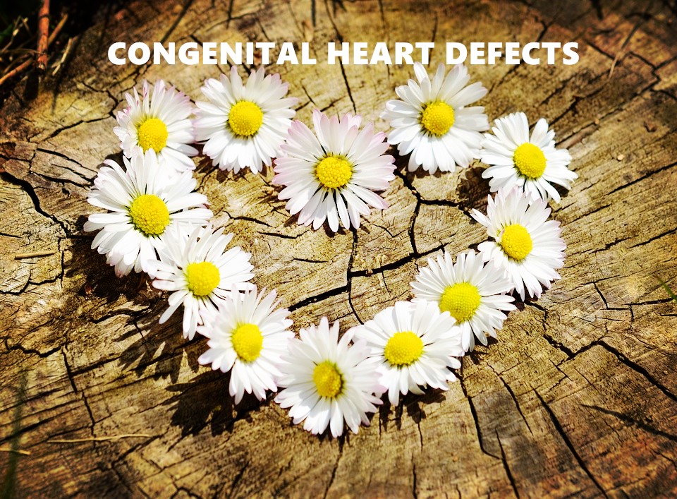
Congenital heart defects - are all valve and septal defects that occurred in the womb before the actual birth . But also the congenital vascular disorders of the heart are among the classic heart defects. The incidence of congenital heart disease is relatively high at about one percent of newborns.
Frequencies of individual heart defects However, the incidence of individual heart defects is much higher, as the following examples illustrate:
31% ventricular septal defect
5 - 8% aortic coarctation
7% atrial septal defect
7% persistent ductus arteriosus
7% pulmonary valve stenosis
3 - 6% aortic valve stenosis
5.5% Fallot tetralogy
In atrial septal defect , the septum between the right and left atrium of the heart is not closed even after birth. Since there is an overpressure in the left atrium, oxygen-rich blood also enters the right atrium. Of course, there is also an atrial septal defect, the so-called ductus botalli. This is trained in all unborn children. It serves primarily as a short circuit defect to circumvent the non-functional pulmonary circulation. In newborns, the ductus botalli is therefore not a congenital heart disease, but is a physiological condition that begins to close after birth. An equally common defect in congenital heart disease is the ventricular septal defect . The dividing wall between the right and left ventricles is not closed, so that blood from the left into the right ventricle presses. Depending on the size of the defect, oxygen deficiency or shortness of breath may occur. Other congenital heart defects are usually associated with larger blood vessels leaving the heart. For example, the aorta and pulmonary artery may be reversed in origin. As a result, only oxygen-poor blood enters the body, which is incompatible with life. Stenoses (constrictions) in the area of the pulmonary valves or the aortic arch are also quite common. In the so-called Fallot tetralogy , four groups of heart defects occur simultaneously - a ventricular septal defect, a pulmonary valve stenosis, an enlargement of the right heart and an aortic anomaly. In general, the more severe the heart defect, the more likely heart surgery is to be the only remaining therapy. Some heart defects in detail A heart defect is usually not necessarily discovered at birth. More often symptoms occur later in life. Only in some cases are the symptoms so serious that the heart defect can be detected even before birth or within the first few weeks of life. Then usually the pulmonary artery and the pulmonary valve are affected. Blood flow from the right ventricle into the lungs is affected by heart failure and symptoms of hypoxia can occur. a) Pulmonary atresia In this type of heart defect, the three flaps of the sails do not open or are not formed. As a result, the blood can not flow from the right ventricle into the pulmonary artery. The blood does not flow through the lungs and can not be oxygenated. b) Pulmonary valve stenosis Pulmonary valve stenosis is also a defect in the valve leaflets of the pulmonary valve. This opens only incompletely in cardiac activity, whereby the outflow path of the blood is restricted. Through this narrowing, the heart has to build up a higher pressure to pump blood into the lungs. c) The Fallot tetralogy This clinical picture of congenital heart disease is very complex. It actually consists of four different congenital heart defects that occur simultaneously. First, this is a pronounced pulmonary valve stenosis, a ventricular septal defect - a hole in the muscle wall between the left and right ventricles. The overpressure in the right ventricle due to pulmonary valve stenosis constantly forces blood through the ventricular septal defect. The resulting low-oxygen mixed blood leads to the formation of oxygen deficiency symptoms in the systemic circulation. In addition, Fallot tetralogy has an aortic abnormality that can affect blood transport from the heart. d) The transposition of the large cardiac vessels In 5% of all cases, a very serious congenital heart defect occurs - the so-called transposition of the large cardiac vessels. This refers to a faulty connection of the aorta and pulmonary artery to the heart chambers. The aorta arises here in the right ventricle. At the same time, the pulmonary artery is discharged from the left ventricle. As a result, no oxygen-rich blood enters the systemic circulation; a deadly constellation that requires immediate postnatal surgery to save the newborn. Defects of the heart septum Frequently children are born, who have a heart defect in the area of the heart septum. These may be smaller or larger wall defects that cause mixed blood in the affected atria or ventricles. Mixed blood arises because oxygen deficient blood from the body is mixed with oxygen-rich blood from the lungs via the defect. The result is blood with a lower oxygen content than necessary. Depending on the size of the septal defect, symptoms of different severity occur. If the hole is very large, it produces very low-oxygen mixed blood and the oxygen supply of the body suffers. This can be recognized by an altered, rather bluish skin coloration and an increasingly lower load capacity of the child. In these cases, only pediatric cardiac surgery can help to close the defect through surgery. However, smaller defects usually remain unrecognized for years due to their weaker symptoms. Very often, heart defects are detected via the ECG, cardiac catheter or other imaging techniques. The doctor will discuss the best course of action with you as the parent of the affected child. In addition, not every heart defect must be operated on the same. Often, it is sufficient to first check smaller septum defects regularly by means of ECG. Especially in infancy or childhood, many holes in the heart septum between the right and left half of the heart close by themselves. Fortunately, surgery is usually superfluous. However, if a hole persists later and such a heart defect is not treated, threaten sometimes serious complications, such as inflammation, cardiac arrhythmia, valvular heart disease or permanent lung damage. Congenital heart defects in adolescents If a child grows up in the course of his or her youth, combinations of an already corrected congenital heart defect and newly acquired heart defects can occur. Therefore, it is also not uncommon that operators later undergo a heart surgery again. To reduce the risk of scars and repeated stress on the child's body and mind, today, as a rule, all atrial septal surgeries can be performed as a minimally invasive procedure. If severe heart defects are treated as early as infancy, studies have also shown that children then continue to develop normally. Symptoms of congenital heart disease A whole range of symptoms may be evidence of congenital heart disease. Often the pediatrician is the first port of call when these symptoms occur. But how can you identify a possible congenital heart defect in your child? The main cause of the occurrence of symptoms is the increasing lack of oxygen. This becomes visible on the basis of the cyanosis (blue coloring) of skin, lips and nail beds. In addition, however, other symptoms such an accelerated or difficult breathing, listlessness and pale, damp cold skin occur. Also, shortness of breath, tiredness, tachycardia and swelling of feet, ankles or stomach occasionally occur in a congenital heart defect. Diagnosis and therapy of congenital heart defects The entire spectrum of congenital heart defects ranges from the very simple heart defect, which affects the cardiovascular system only slightly, to the very serious heart defect, which leads to an early death without suitable therapy. Overall, no normal life expectancy can be achieved without surgical therapy for moderate and severe congenital heart disease. Due to the improved modern diagnostic methods, many of the heart defects are already recognizable within the first year of life. However, particularly severe heart defects associated with poorer oxygenation are very distressing to the child after birth and require rapid treatment. The prenatal diagnosis makes it possible today to diagnose congenital heart and vascular malformations very early. Many of the severe heart defects are already diagnosed prenatal, ie prenatal. The antenatal diagnosis is not there to be able to schedule an early abortion in severe congenital heart defect. Rather, an optimal care for the newborn after birth is to be made possible. Many congenital heart defects cause loud heart sounds as the blood flow is swirled or shorted due to narrowing or broken heart valves. Such heart sounds can be detected very easily by stethoscope. Depending on the type of noise, its origin can be determined. Also important for the diagnosis of congenital heart defects is the electrocardiogram, also abbreviated as ECG. Through the derivation of the cardiac currents, the doctor can infer the size and position of the heart and, above all, cardiac arrhythmias. However, the most important diagnostic method today is echocardiography. This ultrasound examination shows the heart in all its structures very accurately. Thus, almost all heart defects are visible. In addition, the heart function can be assessed and the condition of the individual heart components can be estimated. This examination method is used for any suspected congenital heart disease. It is completely painless and without risk and is therefore used as a very gentle procedure in children. Further, usually much more specialized investigations are used depending on the type of suspicion. In order to determine a congenital heart defect more accurately, there is the possibility of cardiac catheterization, at the same time an intervention can be done on a heart valve. Other imaging techniques include magnetic resonance imaging (MRI / MRI) and computed tomography (CT). All intervention for the purpose of therapy, both surgical cardiac surgery and interventional with cardiac catheter, aim to close the congenital heart defects (holes, shunts). At the same time, constrictions, so-called stenoses, can be treated or heart valves can be repaired. Thus, the full or at least a gradual capacity of the diseased heart is restored. Severe heart defects in the operating room For very severe heart defects, a simple corrective surgery is often impossible. In this case, several steps must be taken to stabilize the patient and improve his quality of life. The highest priority is ensuring body and lung circulation. Usually, doctors create artificial connections to create mixed blood, so that at least a minimum supply of oxygen is guaranteed. It is interesting that the heart can be bypassed. Low-oxygen blood is passed directly from the large body veins into the pulmonary artery and there enriched with oxygen. This relief of the heart may improve blood flow, which may improve cardiac arrhythmias and thus increase the quality of life. Transposition of the large cardiac vessels A particular challenge among the severe congenital heart defects is the transposition of the large vessels of the heart. The artery leading to the lungs is located at the site of the aorta in these children and the aorta in turn branches off into the lungs. This makes it virtually impossible for oxygen-rich blood to enter the body. Without the rescue operation, these newborns die very soon after birth. The oxygen exchange takes place in the first days of life only via so-called postnatal shunt openings. Therefore, surgery must be performed within a few days after birth. The large body artery and the pulmonary artery are loosened in this OP of the heart, exchanged against each other and connected in their correct position with the heart again. Is there a precaution for congenital heart defects? In fact, there are a number of known risk factors that can have a damaging effect on the developing heart. It is therefore important to avoid these risk factors in the first place. Especially girls should be vaccinated against rubella so that they do not fall ill during a later pregnancy. If you also need medication during pregnancy, you should consult a doctor before taking it. Among the risky products are mainly over-the-counter medications and vitamin tablets. Alcohol and nicotine forbid themselves during and after pregnancy (lactation). Especially important for expectant mothers is participation in all prenatal examinations. During these regular inspections, a congenital heart defect can be detected during pregnancy, ie before birth. For this purpose, the baby's heart is examined more closely during pregnancy with ultrasound. The more skilled the doctor in this case and the better the ultrasound device used, the higher the likelihood that a possible heart defect will be detected safely.
Congenital heart defects - are understood to mean various diseases that occur from birth through a disruption of the development of the heart . They can go unnoticed for years after birth or, depending on their severity, they can be noticed immediately or even lead to death. In more than 90 percent of congenital heart defects, the cause is unknown. They can occur singly, in combination or as part of other illnesses.
Frequency of congenital heart defects Out of 1,000 live births, 8 to 10 children have a congenital heart defect or a large vessel defect that leads to the heart . Congenital heart defects are thus the most common congenital diseases. Without treatment , 25 percent of children die in infancy and around 60 percent die in their first year of life. Only 15 percent reach adulthood without treatment. 25 percent of children still suffer from other illnesses , such as a number of chromosomal defects (genetic diseases). As an example, the Down syndrome is mentioned, which is associated to 30 - 40 percent with narrowing of the main artery ( Aortenisthmusstenose ). Classification and presentation of possible congenital heart defects Congenital heart defects can be divided into the following groups:
Cardiac and vascular defect without short-circuit connection, with pressure load of the ventricle (eg aortic coarctation, aortic stenosis, pulmonary stenosis)
Malformation with pulmonary circulation or volume load of the small circulation: Left, right shunt (eg ductus botalli, ventricular septal defect, atrial septal defect)
Malformation with pulmonary blood flow: right, left shunt (eg, Fallot's tetralogy, ventricular septal defect)
Rotational malformations, which are often combined with atrial or ventricular septal defects and pulmonary or sub-circulation (eg, transposition of the large arteries, double-outlet left / right ventricles)
Below are explanations of some of the known congenital heart defects. aortic stenosis In the case of aortic stenosis, an adhesion of the pocket flaps usually leads to a reduced opening area . Only by increasing left ventricular muscle mass can the increased resistance at the heart valve be overcome and the heart defect compensated. If the children develop discomfort such as breathlessness or paleness on exertion, the operation serves to restore normal valve function.
pulmonary stenosis A narrowing in the area of the pulmonary valve leads to an increased load on the right ventricle. Often there is a fusion of the pockets which prevents complete opening of the pulmonary valve. In addition to the expansion by means of a cardiac catheter, various surgical procedures for correcting the stenosis can be used, depending on the anatomical findings. The age of surgery depends on the severity of pulmonary stenosis. Tetralogy of Fallot Fallot tetralogy is a cyanotic heart defect that involves the combination of four related cardiac abnormalities : pulmonary valve stenosis, with concomitant increase in right ventricular muscle mass, aortic root displacement, ventricular septal defect. The resulting reduced pulmonary blood flow characterizes the clinical picture with heavy breathing and blue-bloodedness. In the foreground of the always operational procedure is the lung perfusion to be improved. The aim is a complete correction with elimination of pulmonary valve stenosis and closure of the ventricular septal defect in infancy. However, this is not always possible due to a complex change in heart anatomy or heavy blue crops. For improved pulmonary blood flow, a short circuit of the aorta with the pulmonary circulation is initially created. A correction will be made later. Open ductus arteriosus botalli (ductus botalli) The Ductus Botalli connects the Pulmonalarterienstrombahn with the main artery and leads before birth, the blood coming from the right ventricle to the systemic circulation. This bypass of the pulmonary circulation is, among other things, important for the oxygen and nutrient supply of the unborn child. Normally, the ductus botalli closes spontaneously in the first days after birth. However, if this occlusion fails, more blood enters the pulmonary circulation in the opposite direction. Left ventricular strain and pulmonary hypertension are the result. The focus of treatment is the occlusion of the ductus botalli. An operative ligation occurs occasionally already in the newborn age .
Transposition of the Great Arteries (TGA) In this severe cyanotic heart defect, the aorta originates from the right ventricle and the pulmonary artery from the left ventricle. Survival is only possible if there is a sufficient connection between the pulmonary circulation and the systemic circulation. Due to the pronounced cyanosis after birth, an operation to transpose these two large arteries and the coronary arteries is required within the first few weeks of life . Ventricular septal defect (VSD) Ventricular septal defect (VSD) is the most common congenital heart defect . Due to an opening in the ventricular septum, arterial blood increasingly passes from the left ventricle into the right ventricle. Depending on the size of the ventricular septal defect, hypertension in the pulmonary circulation develops in addition to the left heart strain. If spontaneous occlusion does not occur in the first years of life, surgical correction of the ventricular septal defect is required. The closure of the opening in the septum by means of seam or art patches. Atrial septal defect (atrial septal defect, ASD) In atrial septal defect, a defect in the atrial septum redirects some of the blood from the left atrium back to the right atrium . This results depending on the size of the atrial septal defect an increasingly circulating pulmonary circulation with concomitant right heart strain. If the pendulum volume exceeds a critical value, closure of the defect is required. In large atrial septal defects and unfavorable position, the opening is surgically closed mostly in preschool age. Causes of congenital heart disease If there is growth disturbance in the area of the heart and / or the large supply and / or discharge vessels during cardiac development in the womb, congenital heart defects result. In more than 90 percent, the cause is unknown. One cause may be infectious diseases , for example, redness, herpes or cytomegalovirus infection, in the first trimester of pregnancy. In addition to alcohol consumption , drug intake can also influence embryonic heart development. Genetic radiation damage can also lead to congenital heart defects. Symptoms vary Symptoms may vary depending on the heart defect. The most noticeable is a heavy cyanosis (Blauchucht). With incipient heart failure, the body can no longer be sufficiently supplied with oxygen. Children and adults with congenital heart disease are constantly tired and find every activity strenuous. They breathe fast and short. Cardiac arrhythmias can also be the result of congenital heart disease. In adulthood, surgically corrected or not, damage is most likely to be noticeable with typical signs of heart failure. In addition to tiredness and fatigue, these are shortness of breath and cardiac arrhythmia. Diagnosis of a congenital heart defect As a drug of choice (gold standard), the ultrasound of the heart ( echocardiography ) has proven itself. In children, echocardiography through the chest wall is usually sufficient from the outside. Increasing chest circumference and rib strength in adults with complex heart defects is indicated by " swallowing ultrasound" (TEE), where a small ultrasound head is inserted into the esophagus via a tube and the doctor has an unobstructed view of the heart from behind the esophagus allowed. Furthermore, examinations using computer and magnetic resonance tomography are available. As an invasive method, with puncture of the vessels in the groin, both the small and the large systemic circulation can be visualized and assessed by means of contrast media (angiography). Protection against inflammation of the inner lining of the vessel Both children and adults with congenital heart defects have to be saved for life from developing inflammation of the endocrine (endocarditis). Most commonly, the heart valves are affected. In particular with interventions at the dentist, in the mouth and throat area, gastrointestinal tract as well as at the urinary tract a suitable "Endokarditisprophylaxe" must be operated. Treatment of malformation Corrective surgery has been developed over the past 25 to 30 years for almost all cardiac and vascular malformations . In a few cases, the operation will create a completely healthy heart. In most cases these are "repair conditions" with critical points in the area of the seams. The development of pediatric surgery has made it possible, especially in the last 10 to 15 years, to successfully operate on complex congenital heart defects. Due to the short surgical history, long-term results will not be available in the next few years. Furthermore, in recent years, catheter techniques have been developed which allow to treat heart septum defects and valve narrowings. Patients with corrected congenital heart disease that have outgrown adolescence are being treated by pediatric and adult cardiologists. Prognosis for children with congenital heart disease
The prospects of children with congenital heart disease are very different, depending on the severity of the heart failure and the time of diagnosis. With medical advances, especially cardiac surgery, 90 percent of children with congenital heart disease reach adulthood. In specialist centers, pediatric and adult cardiologists have joined forces to ensure optimal care for patients with congenital heart disease. Although adolescents and adults with congenital heart disease are chronically ill, they can achieve a good quality of life through targeted care and counseling.
In pregnancy - it can cause high blood pressure occur, which can be dangerous untreated for mother and child. It can become a deadly threat to mother and unborn child: preeclampsia. Your symptoms and how the doctor treats you.
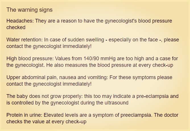
Preeclampsia, under this term, probably the least pregnant women can spontaneously imagine something. The colloquial term "pregnancy poisoning" suggests: It is a serious complication - in the worst case, it can end fatally. The causes of the disease, which was formerly also called EPH-gestosis and affects about two to three percent of all pregnant women , are still unclear. The symptoms are often nonspecific, making the diagnosis difficult. Depending on the degree of severity, the medicine distinguishes four forms of the so-called hypertensive pregnancy disorder, whose leading symptom is the high blood pressure: - Gestational hypertension - Preeclampsia (pregnancy poisoning) - eclampsia - HELLP syndrome If left untreated, hypertensive pregnancy can be very dangerous for both mother and child. Failure to implant the fertilized ice into the uterus causes changes in the blood vessels in the developing placenta . This leads to the release of signaling substances from the altered placenta into the circulation, which then trigger the symptoms. If left untreated, it can lead to life-threatening blood pressure crises in the mother, to miscarriages or damage to the child. Causes of high blood pressure during pregnancy What causes a hypertensive pregnancy is not yet fully understood. However, everything indicates that heredity plays a role: if the mother was suffering from high blood pressure during pregnancy, the risk of her daughter also being affected is increased by about 25 percent. Even if the mother of the child's father had it, the risk can be increased. Apparently, among other things, the genes of the unborn child are responsible, if the mother develops a gestational pressure. Substances of the misfolded placenta enter the mother's bloodstream and cause high blood pressure and also a blood coagulation disordercan develop. The clinical picture that emerges can vary in severity - from pregnancy hypertension to preeclampsia and eclampsia to HELLP syndrome. Of these hypertensive diseases, which develop only during pregnancy, a distinction is made between "high blood pressure during pregnancy". These are patients who already had high blood pressure before pregnancy. In most cases, sufficient therapy with uncomplicated pregnancies is expected here. However, the hypertension remains after pregnancy and must continue to be treated. Symptoms of pregnancy hypertension The ideal blood pressure is 120/80 mmHg. Values starting at 140/90 mmHg are considered elevated blood pressure. Severe hypertension is present when the lower (diastolic) value is 120 mmHg or above. In women who had low blood pressure before they were pregnant , the values shift slightly downwards - what is considered normal in other women can be considered as elevated blood pressure. Symptoms of pregnancy hypertension The affected women usually do not notice the onset of pregnancy high pressure. At the preventive appointments , however, the doctor determines the increased blood pressure. Increased scores are not always worrying because, for example, blood pressure also increases when you're excited. Higher values - especially after the 20th week of pregnancy - are always a reason for regular blood pressure checks. Pregnancy-related hypertension returns to normal about three months after birth . Symptoms of preeclampsia (EPH-gestosis, pregnancy poisoning) A pre-eclampsia can also go unnoticed at first. At the latest at the check-up then it is noticeable that in addition to the increased blood pressure, protein in the urine can be detected. The patient may also notice water retention in the feet and lower legs, hands, and facial area. Other symptoms that should alert you are : - Headache , dizziness , flicker in front of the eyes - photosensitivity - drowsiness - confusion - Pain in the upper abdomen If you experience one or more of these symptoms, contact your doctor quickly. Symptoms of Eclampsia Eclampsia is a serious complication of pre-eclampsia. In addition to pre-eclampsia symptoms, seizures occur before, during and after birth. Before the seizure, sufferers often complain of headaches, blurred vision and upper abdominal pain. Symptoms of HELLP syndrome HELLP syndrome is also a serious complication of pre-eclampsia. HELLP is the main symptoms of this disease: Hypertonie (high blood pressure), e levated li ver enzymes (elevated liver enzymes) and low p latelet count (reduced number of platelets , i.e. platelets). Symptoms of HELLP syndrome usually do not occur until the 25th to 30th week of pregnancy: In addition to the signs of preeclampsia, there are strong, usually radiating upper abdominal pain due to liver damage: For many patients, it feels as though they are a ring compressed below the costal arch. Treatment of high blood pressure in pregnancy The easiest way to treat pregnancy high pressure and possible complications such as pre-eclampsia is to recognize and treat the blood pressure increase at an early stage. Almost always, you need medication that lowers your blood pressure. Your doctor knows which ones are suitable for pregnant women. In severe forms of hypertensive pregnancy, premature birth may be required - for example, when the mother has seizures (eclampsia), threatens kidney failure, or indicates that the unborn child is no longer receiving adequate care. Symptoms usually from the 20th week of pregnancy One thing is certain: "The placenta does not nest in the eighth to thirteenth week, as a result of which harmful substances enter the maternal circulation", explains Dr. med. Dietmar Schlembach, Chief Physician of the Clinic for Obstetrics at Vivantes Klinikum Neukölln in Berlin. "They have a delayed, negative effect on the mother's vascular system, probably as soon as a certain threshold is exceeded." From about the 20th week of pregnancy , the first signs can show. Depending on their severity, they range from mild headaches to fluid retention in the tissue to severe upper abdominal pain and vomiting. Close monitoring According to the definition, only two criteria have to be fulfilled for pre-eclampsia: hypertension and increased protein levels in the urine. "The actual clinical spectrum is very broad," Dr. Stefan Verlohren, Senior Physician of the Clinic for Obstetrics at the Berlin Charité. If the clinical picture is limited to increased protein levels in the urine and increased blood pressure (from 140/90 mmHg), the gynecologist usually monitors the pregnant women more closely. Partly he prescribes antihypertensive agents. Eclampsia: complication with seizures Heavier gradients always require a hospital stay. "In eclampsia, for example, seizures occur during pregnancy, during or after birth , which can be life-threatening for both the pregnant woman and the child," says Verlohren. For example, placental detachment or kidney failure is possible. "In rare cases, the blood pressure very quickly rises very high, so that it can lead to circulatory disturbances in the brain to stroke in pregnant women," said Schlembach. HELLP syndrome: impaired liver function HELLP syndrome is another serious complication. Here are disorders of liver function and blood clotting, sometimes without the blood pressure increases. Upper abdominal pain is a warning sign. "Therefore, any pregnant woman should visit the gynecologist for pain in the upper abdomen or behind the sternum, in case of sudden swelling especially in the face, with very rapid weight gain or nausea and vomiting immediately and not go to the family doctor." Clear prognosis now possible Up to now, it has been virtually impossible to predict the presence of pre-eclampsia in the first suspected symptoms. An international study has now shown that the ratio of certain protein messenger substances in the blood reliably excludes or predicts possible gestosis. It's about a certain separation value. Stefan Verlohren, senior author of the study: "Women who are below the age of 38 do not contract pre-eclampsia within one week with 99.3 percent confidence." This does not sound spectacular to laymen, but offers those affected a great relief. You do not have to worry about a sudden deterioration of your condition in the short term. Instead of being admitted in a hospital, they are allowed to look after themselves at home. Early preeclampsia particularly problematic The earlier signs of preeclampsia appear, the more problematic for both mother and child. The actual therapy consists in the timely delivery and the removal of the nut cake. Only then does the mother's vascular system recover completely under normal circumstances. "That puts us at an early eclampsia in the 24th week a dilemma: How can we extend the pregnancy as far as possible to give the child more time to develop without harming mother and child? "Schlembach, the chief physician, says that if the blood pressure can be stopped by medication and the child is not in need of care, they become pregnant Mostly monitored intensively to gain a few more weeks, here the new blood test, which is still not a cash benefit, helps doctors and affected women to better prepare for impending complications. New treatment method is being researched A new therapeutic approach, which is currently used in only a few centers and under a strict study protocol, is the so-called blood wash. "The problematic messenger, the protein sFlt-1, is purposefully removed from the blood," says Verlohren. "The method is still in the experimental stage and is currently only in individual cases in question." A quick cure is not yet available. Until then, women should stay alert for early warning signs and know if they belong to a high risk group. Know risk factors "Especially young first-time mothers are more likely to develop pre-eclampsia, but even with multiple pregnancies or after fertility treatments there is a higher risk," says Schlembach. Existing autoimmune diseases, diabetes, high blood pressure or other previous cardiovascular problems are also a reason for pregnant women to ask the doctor at the slightest suspicion for advice. Although women who have already suffered from pre-eclampsia have a slightly higher risk of developing another pregnancy, the symptoms can also be completely absent.
What you can do yourself If you have been diagnosed with pregnancy hypertension, you are likely to be on medication to lower your blood pressure. If you suffer from water retention, it is better not to take medicines or teas that have a flushing effect - unless your doctor has prescribed it. The advice to consume low salt to promote the elimination of fluid is outdated. Eat well-balanced and healthy , and save yourself otherwise - a lot of rest, no stress and no excitement. NON-MEDICAMENTAL MEASURES Pregnant women with high blood pressure should take care. Strict bed rest is rarely required. It has no favorable influence on the course of preeclampsia. Low-salt food is not recommended. Salt deprivation lowers the plasma volume, which is in any case lowered in hypertensive pregnant women. Also from low-calorie diets for weight loss is not recommended during pregnancy. Women with preeclampsia should be cared for inpatient care. INDICATION AND GOAL MEDICINE THERAPY Whether pregnant women with mild hypertension benefit from a drug therapy, remains open. An American consensus report in 1990 recommended treating diastolic pressures of 100 mm Hg and above. Other authors consider antihypertensives to be indicated only at 105 to 110 mm Hg diastolic. In order not to endanger the blood supply of the fetal sera unit, the pressure must not be lowered too much. For pre-eclampsia, diastolic values of 100 mm Hg would be desirable. In women with renal disease or left ventricular hypertrophy, values above 90 to 95 mm Hg should be avoided. The treatment is intended to prevent organ complications such as brain haemorrhage in the mother. The therapeutic goal is derived from high-pressure treatment in the general population and from retrospective studies with pregnant women. There is insufficient evidence that the available antihypertensives may favorably affect the blood supply of the fruit and fetal growth, prevent pre-eclampsia, prevent the worsening of pre-eclampsia or premature birth. SELECTION OF HIGH-PRESSURE MEDICINAL PRODUCTS Most experiences and randomized studies on chronic hypertension in pregnant women are available with the central alpha- blocker methyldopa (PRESINOL et al.), Which is only rarely used in general high-pressure treatment. Fruit damaging effects are not described. Methyldopa is the only extreme pressure agent whose potential long-term effects on children have been studied in studies. There are no undesirable effects until the age of seven. However, users of methyldopa often suffer from fatigue, dizziness and dry mouth. The agent should be avoided in the case of a history of depression. Threats such as hemolytic anemia, hepatitis, vasculitis and lupus erythematosus-like disorders are to be expected. The direct COOMBS test is positive for 20% of users. Nevertheless, methyldopa is still considered the drug of choice for chronic hypertension in pregnant women or for long-term treatment of pre-eclampsia, especially in English-speaking countries. Proliferation in high-pressure therapy Pregnant women have beta-blockers . Teratogenic effects in humans are not described. In the newborn, treatment of the mother may cause bradycardia, hypotension, hypoglycemia and respiratory depression. Obstetricians and pediatricians should be informed about the medication. The child should be closely monitored. Internationally, different beta-blockers are preferred. However, it has not yet been proven whether certain active substances or groups of active substances actually have advantages. German textbooks recommend beta1-selective receptor blockers. Studies do not justify this preference. The relatively cardioselective atenolol (TENORMIN et al.) Reduces uteroplacental perfusion compared to nonselective pindolol (VISKEN et al.). From the second trimester onwards, the weight of the newborn decreases compared to placebo. Metoprolol (BELOC et al.) And Atenolol attenuate the fetal heart rate. Compared with the calcium antagonist nicardipine (ANTAGONIL), metoprolol decreases the perfusion of the fetal serine unit. The number of cesarean deliveries increases due to fetal distress. However, one of our consultants notes that metoprolol is widely prescribed in practice without any indication of serious adverse effects from individual reports or reports to the European network of teratology consultation centers (ENTIS). However, caution seems appropriate when fete retardation is to be feared, as in pre-eclampsia, as well as during long-term use in pregnancy. Atenolol is not recommended. This also applies to propranolol (DOCITON and others). The non-selective beta-blocker is associated, among other things, with low birth weight and increased perinatal mortality. A Scandinavian workshop recommends the non-selective beta-blocker pindolol , which is said to reduce heart rate and stroke volume because of intrinsic stimulating activity (ISA). The blocker, which is little used here in Germany, does not appear to influence the fetus unfavorably according to previous findings. However, larger studies with pindolol and data on long-term use during pregnancy are lacking. In English-speaking and Nordic countries also obsolete in Germany alpha and beta adrenergic receptor blocker is labetalol (Switzerland: Trandate) is recommended. After taking in chronic hypertension from the end of the first trimester there is no difference to methyldopa. However, after use in pre-eclampsia, one study found a reduced neonatal weight over the control group, which is ingested only. Dihydralazine (NEPRESOL et al.), In other countries hydralazine (in TREPRESS), is mainly used for the acute parenteral treatment of severe hypertension during pregnancy, especially in preeclampsia. Based on their extensive experience, these vasodilators are still considered the drug of choice. In order to avoid a sharp drop in blood pressure with worsening of the uteroplacental blood flow, it is necessary to dose very slowly or perfusor-controlled. Because of mutagenic effects, (di-) hydralazine should not be used in the first trimester. There is no evidence of teratogenic effects in humans when dihydralazine is taken during organogenesis. The long-term use per os is less common for pregnant women today because of frequent parasitic effects. Headache and vomiting can mimic threatening eclampsia. Increase in uterine and placental blood flow in dihydralazine seen in previous studies was later confirmed. calcium antagonists are not suitable for the first trimester of pregnancy due to teratogenic effects in animal experiments and inadequate documentation in humans. Nifedipine (ADALAT et al.) Is among the best studied . The uteroplacental blood flow does not appear to attenuate the calcium antagonist. In the acute treatment of severe hypertension, non-sustained nifedipine performs similarly in several studies as (di-) hydralazine parenterally. However, nifedipine capsules are no longer recommended during high-pressure crises because of poor control of the drop in blood pressure with potentially life-threatening cardiovascular consequences. For prolonged use of dihydropyridines retardation forms should be used. Caution is advised in women with pre-eclampsia, who get magnesium for the prevention of convulsions: Simultaneous intake of calcium antagonists can provoke a sharp fall in blood pressure1 (including constipation to the ileus). Long-term data for the treatment of chronic hypertension with retarded nifedipine are missing. Not least on suspicion of carcinogenicity, we consider calcium antagonists as a reserve during pregnancy. CONTESTED AND CONTRAINDICATED MEDICINES Diuretics are non-teratogenic. Theoretical concerns - the reduction in plasma volume - argue against their application. A meta-analysis of randomized trials involving more than 7,000 women showed no adverse effects on the fetus 10 years ago. Current data is missing. If the blood pressure in chronic hypertension is well adjusted prior to pregnancy with diuretics, the medication may be maintained. There are rarely any reasons for recruiting during pregnancy (eg pulmonary edema). For pre-eclampsia and intrauterine growth delay, diuretics are considered contraindicated. Because of oligohydramnios, kidney failure in neonates and congenital malformations ACE inhibitors prohibit pregnancy. Women who become pregnant on receipt should be promptly switched to other high-pressure products. Based on experience with ACE inhibitors, angiotensin II antagonists are considered contraindicated. The data on alpha-receptor blockers is far from sufficient for recommendations. Following minor studies and meta-analyzes, calcium (CALCIUM SANDOZ et al.) Was orally suggested for the prevention of pre-eclampsia. However, in a randomized clinical trial of more than 4,500 healthy first-time mothers, 2 g of calcium daily are not protective against either pregnancy hypertension or pre-eclampsia or obstetric complications. Also the prophylaxis with Acetylsalicylic acid (ASPIRIN et al.) Fails in two large randomized trials. Magnesium per os (MAGNESIUM VERLA and others) is also not recommended for protection against preeclampsia. In manifest eclampsia but magnesium sulfate (serving (MG 5 SULPHATE etc.) parenterally peripheral attenuation of seizures CONCLUSIONS In the high-pressure treatment of pregnant women, two otherwise rarely used veterinary medicinal products are preferred: methyldopa (PRESINOL et al.) Per os for long-term treatment and dihydralazine (NEPRESOL et al.) Iv for acute therapy. The proven methyldopa for fetal development has the disadvantage of frequent and - although rarely - possibly threatening disturbances for the mother. Conflicting recommendations are available on the more tolerable beta-blockers for pregnant women. Data on the influence on uteroplacental blood flow and fetal development are inconsistent or absent. Short-term use of metoprolol (BELOC, etc.) seems safe. A Scandinavian working group recommends pindolol (VISKEN et al.). Atenolol (TENORMIN etc.) is not recommended. For the long-term use in chronic hypertension, the reserve for methyldopa is also the relatively well studied alpha and beta-blocker Labetalol (Switzerland: TRANDATE). A teratogenic effect has not yet been demonstrated in any of the antihypertensives commonly used today, so that accidental ingestion is not an indication for termination of pregnancy.